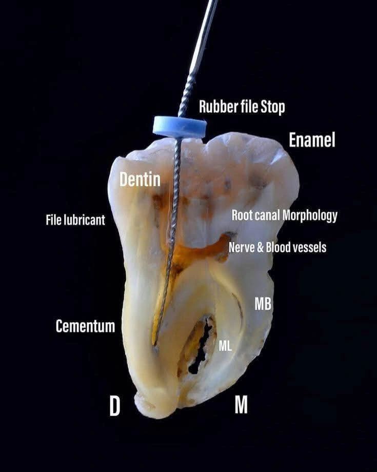
Every tooth tells a story — and this image is a perfect example.
It shows the entire anatomy of a tooth during a root canal procedure – from enamel and dentin to the nerve & blood vessel system we clean and shape.
🛠 What you see here:
✅ Rubber File Stop – for precision length control
✅ Dentin & Enamel – the tooth’s protective layers
✅ Root Canal Morphology – the pathways we clean
✅ Cementum – anchoring the tooth in place
As a dentist, I find it fascinating that something so small has such complex biology. It reminds me that dentistry is not just about drilling and filling – it’s about preserving life inside teeth and giving patients pain-free smiles.
💡 Key Takeaway:
Root canal treatment is NOT a scary procedure – it’s a science-backed, precision-driven way to save teeth and avoid extractions.